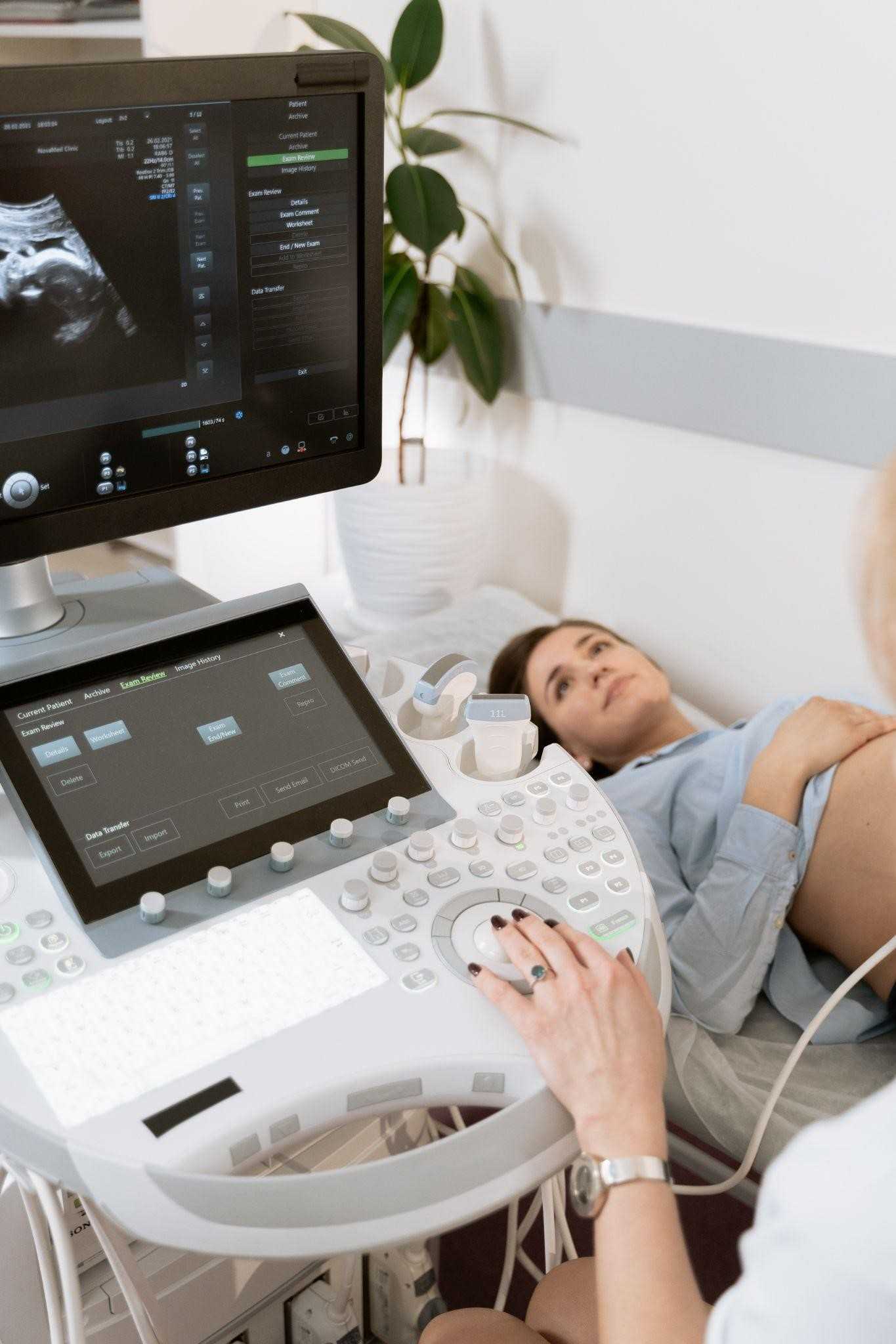
Radiology is commonly referred to as medical imaging, where medical practitioners recreate images of different body parts aiming at diagnosing, treating, or monitoring. Hi-tech imaging provides the market with some of the most superior medical imaging equipment, such as X-Ray equipment, Computerized Tomography (CT) scans, C-Arm machines, and DR panels.
Another widespread type of imagining machine is the ultrasound machine. The device uses sound wave frequency and converts them to an image—the primary use is for diagnostic, testing, or therapeutic reasons. Ultrasound is the safest way to examine internal organs since it does not use radiation. Most of us are familiar with external ultrasound. Still, an internal ultrasound is inserted into the body, and the endoscopic ultrasound passes through the mouth.
The main focus of the following article is the external ultrasound by outlining the importance of ultrasound in pregnancy. Below are the five key benefits of ultrasound during pregnancy.
Ultrasound Verifies Gestational Age
Normal human gestation is commonly considered as 40 weeks gestation period. The human gestation period is usually between 37 weeks to 41 weeks in the medical language. During this period, the fetus undergoes steady growth. An ultrasound test measures the fetus against a time-calibrated chart to monitor development. The mother estimated the fetus’s age by providing the last time they received their menstrual cycle. The doctor can now compare the development and the estimated age to make conclusions on the due date.

Ultrasound Monitors Placenta Problems
The placenta health is responsible for feeding the fetus and supplying the fetus with oxygen. The placenta health is crucial during pregnancy. In the unfortunate instances of the placenta, the placenta may experience some problems that affect its functionality. Ultrasound test helps in identifying placenta problems like;
Placenta Abruption – A condition where the placenta peels off entirely or partially. The situation results in heavy bleeding, and it’s considered an emergency. The condition causes the deprivation of nutrients and oxygen to the fetus.
Placenta Previa – A condition where the placenta covers the cervix, opening the uterus. The condition mainly occurs during the early stages of pregnancy and can be corrected as the pregnancy develops.
Placenta Accreta – In normal circumstances, the placenta dethatches entirely from the uterine wall after childbirth. The condition leaves a part of the placenta attached to the uterine wall due to blood vessels growing deep into the uterine wall—the condition results in heavy bleeding during childbirth.
Retained placenta – a retained placenta is when the placenta is not delivered within 30 minutes after childbirth. The condition mainly occurs when the placenta is trapped within the uterus or still attached to the uterine wall. The condition exposes the patient to life-threatening infections or even blood loss.

Ultrasound Checks Multiple Pregnancies
Pregnancy occurs as a single pregnancy where only one child is born. In other occurrences, there could be twins where two children are born, triplets where three children are born, and quadruplets where four children get delivered on a few occasions. During pregnancy, taking an ultrasound test can quickly identify the number of children to expect when they are due. The ultrasound also helps prevent some preventable complications during the delivery of multiple pregnancies. It also helps in monitoring the growth of each fetus in multiple pregnancies.

Ultrasound Helps Identify Congenital Anomalies
Congenital anomalies are diseases or physical limitations that are present from birth. Some easily notable congenital anomalies are physical disabilities like missing limbs or arms. Some parents may decide to terminate the pregnancy in such occurrences. At the same time, some will be physically and emotionally prepared to handle the birth and raising of a child with special needs. If the congenital anomalies could threaten the mother’s health, the doctor may advise of termination of the pregnancy.

Ultrasound Monitors Fetal Position
The average fetus’s position appears as a C with a curved back, head facing inward, and the legs and the arms are folded very close to the body. The position of the fetus changes with the pregnancy’s growth. The fetus’s positions include;
Occiput Anterior (OA) is the best birth position where the fetus’s head is facing down and the legs up. The face usually faces backward, and the back faces the belly. The position is the best as it eases the birth process.
Occiput Posterior (OP) – The position is a close replica of Occiput Anterior, except that the face does face the belly, and the back faces the back. The position hinders the fetus from tacking the chin down during birth.
Breech position – the fetus has the head facing up and the bottom down at the cervix. There are three types of breech positions: complete, frank, and footling breech.
Oblique Position – The rear condition where the fetus sits diagonally across the womb. The condition poses the risk of umbilical cord compression causing blood flow restriction. The situation is back but has chances of happening.
Transverse position – on this condition, the fetal is in a fetal position, but their shoulder, back, feet and hands are very close to the birth canal. In its occurrence, the mother has to undergo a C-section since turning the fetus can damage the placenta causing excessive bleeding.
To Summarize
An ultrasound creates images of the developing fetus and the mother’s placenta using high-frequency sound waves. Different types of ultrasound used during pregnancy are transvaginal ultrasound, fetal echocardiography, and 3D or 4D ultrasound.


Comments are closed.